New electron microscope sees more than an image
By Bill Steele
The electron microscope, a powerful tool for science, just became even more powerful, with an improvement developed by Cornell physicists. Their electron microscope pixel array detector (EMPAD) yields not just an image, but a wealth of information about the electrons that create the image and, from that, more about the structure of the sample.
“We can extract local strains, tilts, rotations, polarity and even electric and magnetic fields,” explained David Muller, professor of applied and engineering physics, who developed the new device with Sol Gruner, professor of physics, and members of their research groups.
Cornell’s Center for Technology Licensing (CTL) has licensed the invention to FEI, a leading manufacturer of electron microscopes (a division of Thermo Fisher Scientific, which supplies products and services for the life sciences through several brands). FEI expects to complete the commercialization of the design and offer the detector for new and retrofitted electron microscopes this year.
“It’s mind-boggling to contemplate what researchers around the world will discover through this match of Cornell’s deep expertise in detector science with market leader Thermo Fisher Scientific,” said Patrick Govang, technology licensing officer at CTL.
The scientists described their work in the February 2016 issue of the journal Microscopy and Microanalysis.
In the usual scanning transmission electron microscope (STEM), a narrow beam of electrons is fired down through a sample, scanning back and forth to produce an image. A detector underneath reads the varying intensity of electrons coming through and sends a signal that draws an image on a computer screen.
The EMPAD that replaces the usual detector is made up of a 128x128 array of electron-sensitive pixels, each 150 microns (millionths of a meter) square, bonded to an integrated circuit that reads out the signals – somewhat like the array of light-sensitive pixels in the sensor in a digital camera, but not to form an image. Its purpose is to detect the angles at which electrons emerge, as each electron hits a different pixel. The EMPAD is a spinoff of X-ray detectors the physicists have built for X-ray crystallography work at the Cornell High Energy Synchrotron Source (CHESS), and it can work in a similar way to reveal the atomic structure of a sample.
Combined with the focused beam of the electron microscope, the detector allows researchers to build up a “four-dimensional” map of both position and momentum of the electrons as they pass through a sample to reveal the atomic structure and forces inside. The EMPAD is unusual in its speed, sensitivity and wide range of intensities it can record – from detecting a single electron to intense beams containing hundreds of thousands or even a million electrons.
“It would be like taking a photograph of a sunset that showed both details on the surface of the sun and the details of darkest shadows,” Muller explained.

The improvement is also exciting to life scientists because collecting all the scattered electrons makes the instrument much more sensitive, using a less intense exposure to get an image and limiting damage to a living specimen.
“The EMPAD records an image frame in less than a millisecond and can detect from one to a million primary electrons per pixel, per image frame,” Muller explained. “This is 1,000 times the dynamic range, and 100 times the speed of conventional electron image sensors.”
“Now we can get a better look at processes inside intact cells,” said Lena Kourkoutis, assistant professor of applied and engineering physics. The low dose of radiation allows multiple exposures, to take time-lapse “movies” of cellular processes or to view the same specimen from different angles to get a clearer 3-D image. Kourkoutis plans to use these techniques in work with the new Cornell Center for the Physics of Cancer Metabolism, looking at how cancer progresses from cell to cell.
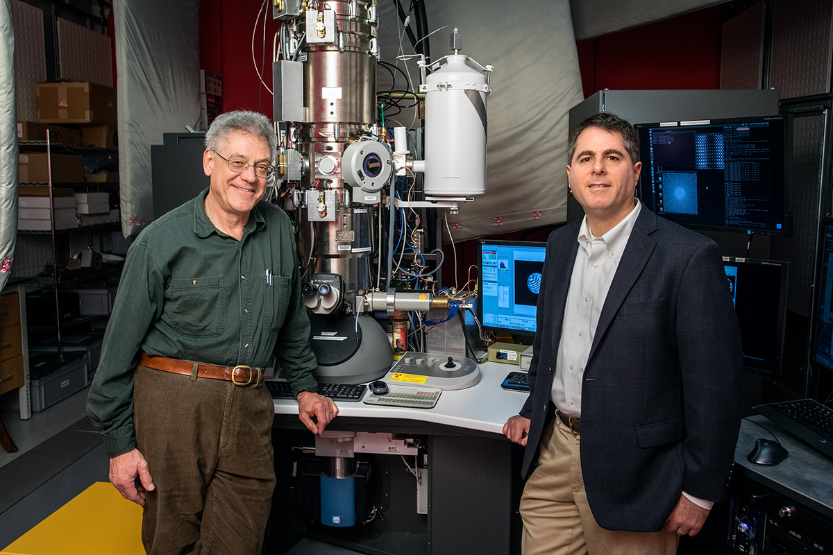
The researchers tested their first EMPAD by installing it in spare port in a state-of-the-art FEI microscope. The prototype is used intensively for experiments in the Cornell Center for Materials Research.
The work is supported by the U.S. Department of Energy. Support also came from the Cornell Center for Materials Research and CHESS, which are supported by the National Science Foundation. The adaptation to the on-campus STEM was supported by the Kavli Institute at Cornell for Nanoscale Science,
Media Contact
Get Cornell news delivered right to your inbox.
Subscribe