Cornell researchers probe secrets of chemical bonding by assembling molecules one at a time
By Bill Steele
Researchers are interested not only in what happens when you combine two chemicals, but also in how the bonds between these chemicals are formed. That understanding might lead to better control over chemical reactions and perhaps even the creation of complex molecules with unusual properties.
Cornell physicists now have shed new light on chemical bonding by combining a single molecule with a single atom to form a new molecule.
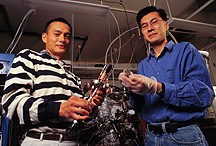
Wilson Ho, Cornell professor of physics, and graduate research assistant Hyojune Lee used a specially designed scanning tunneling microscope (STM) to pick up single molecules of carbon monoxide and graft them onto an iron atom to form molecules of iron carbonyl. By obtaining the new assembly's "vibrational spectrum" -- a measure of the energy in the bonds between atoms -- they verified that a chemical bond had truly been formed to produce a new molecule.
The primary purpose of the work, reported in the Nov. 26, 1999, issue of the journal Science, is to demonstrate a technique for single molecule formation and to learn more about the properties of chemical bonds. But Lee noted that the technology used could have future applications in nanofabrication, the creation of materials and devices at the atomic or molecular level. "One implication is that since we can build up molecules from atoms step by step, in the future it might be possible to build up more complicated molecules from separate ingredients," he said.
The researchers used an STM of exceptional precision to manipulate atoms of iron and molecules of carbon monoxide adsorbed (bonded) on a silver surface in a vacuum. The entire apparatus was cooled to a temperature of 13 degrees Kelvin (13 degrees above absolute zero, or -260 degrees Celsius). The advanced STM built by Ho's research group can resolve parts of atoms and molecules and, by measuring current flow through a single molecule, return characteristic signatures of chemical bonds.
In an STM, a metal tip is suspended a fraction of a nanometer (a nanometer is a billionth of a meter) above a surface and a voltage is applied between the tip and the surface. A tiny electric current called a "tunneling current" flows between the tip and the surface.
The tip is scanned across the surface and at the same time raised and lowered in such a way as to keep the tunneling current constant. The up and down movements are recorded and can be translated by a computer into a relief map of the surface so detailed that it shows the outlines of individual atoms.
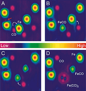
In their recent experiment, the researchers used the STM to scan a surface and locate iron atoms and carbon monoxide molecules. They then lowered the tip over a CO molecule and increased the voltage and current flow of the instrument to pick up the molecule. Finally, they moved the molecule on the tip over an iron atom and reversed the current flow, causing the molecule to bond to the atom and form an iron carbonyl Fe(CO) molecule.
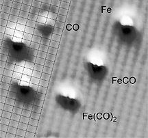
The researchers then added another CO molecule to the Fe(CO), forming a molecule of Fe(CO)2. In subsequent images of the surface, the Fe(CO) molecule appears to have a small lobe on one side, indicating that the CO structure is not attached in a straight up-and-down fashion to the iron atom but is tilted slightly to one side. The Fe(CO)2 molecule appears to have two lobes, indicating that the two CO structures are tilted in opposite directions in a "rabbit-ears" shape. Since there is no way to determine exactly what the angle of the tilt is from their measurements, that would be left to theoretical calculations, the researchers said in their paper.
"By the image change we can predict that a new molecule has been formed, but we need absolute confirmation," Lee said. To measure the vibrational energy of bonds in the molecules, the researchers hold the STM tip at a constant height above a bond and vary the voltage. At certain voltages they will see peaks, where energy is absorbed by the bond. These peaks have a characteristic pattern, or vibrational spectrum, by which the type of bond can be identified. To confirm the presence of a bond between carbon monoxide and iron, the researchers compared the vibrational spectrum of the molecules they had created with the spectra of CO molecules bonded to the silver surface.
In the course of their work, Ho and Lee discovered serendipitously that when a CO molecule was attached to the tip of the STM, the resolution of the instrument increased, enabling the researchers to see the lattice of silver atoms on the working surface. This made it possible to see the bonding sites on the surface for the different chemical species, they said.
The paper in Science is titled "Single Bond Formation and Characterization with a Scanning Tunneling Microscope." The research was funded by the U.S. Department of Energy.
Media Contact
Get Cornell news delivered right to your inbox.
Subscribe


