From nuclear fusion to wiggling ants: Spin-off of energy research produces high-resolution X-ray images of minute objects
By David Brand

The energy that powers the sun would seem to have little in common with the hair on the tip of a housefly's wing. But in a Cornell University lab, the two have found a curious unison.
Using powerful machinery originally developed in the hope of discovering a way to generate energy from hydrogen fusion, scientists in Cornell's Laboratory of Plasma Studies are creating high-resolution images of minute objects, like fly hairs or the fine filaments that keep dandelion seeds afloat in the air.
The machine that produces the images runs a powerful electrical current through a vacuum chamber containing a pair of crossed wires, each many times finer than a human hair. The current is so strong that it causes the wires to explode, forming a plasma -- a dense gas that has become so hot that the atoms in it break down. This plasma is known as the X-pinch, and at the cross point of the wires it is particularly hot and dense. What makes the X-ray images it produces so special is their extremely fine resolution. And, in turn, the very fineness of this detail is being used to determine the size of the X-pinch plasma -- estimated at much less than a thousandth of an inch.

David Hammer, the J.C. Ward Professor of Nuclear Energy Engineering at Cornell and principal investigator in a Department of Energy project to study the properties of the X-pinch, will describe the new imaging technique at a meeting of the American Physical Society, Division of Plasma Physics, being held this week (Oct. 29 to Nov. 2) at the Long Beach Convention Center in Long Beach, Calif. Hammer and his colleagues from the Lebedev Physical Institute, Moscow, Tania Shelkovenko and Sergei Pikuz, will discuss the work in a poster session at 2 p.m. Pacific time, in Exhibit Hall B, and in a lecture Nov. 2 at 9:30 a.m. Pacific time in Room 104BC.
The Cornell lab has been working for the past few years with Sandia National Laboratories, Albuquerque, N.M., to develop an inertial confinement fusion system that uses X-rays. The so-called Z machine that has been built at Sandia is designed to generate an extremely high-power X-ray pulse to create temperatures in the millions of degrees that would result in fusion of the hydrogen fuel. This is the energy source of the sun and other stars, in which hydrogen nuclei combine, or fuse, to produce huge amounts of energy. However, this same research also has yielded the unexpected discovery of X-pinch imaging.
The plasma created by the exploding crossed wires lasts for less than one microsecond, but in that time, it implodes and forms one or two plasma points with temperatures as high as 10 million degrees Celsius that last for less than a billionth of a second. The high-density plasma -- almost the density of a solid -- generates bursts of X-rays that can produce extremely high-resolution radiographs (X-ray photographs) of very small objects.

Because the detail shown in the radiograph is determined by the size of the X-ray source, microscopic details can be shown at very high resolution. Indeed, the plasma points that emit the X-rays are so small that their size is still unknown. "What we're doing now is making little nanofabricated structures, and then we will image those structures and see what the resolution is," says Hammer.
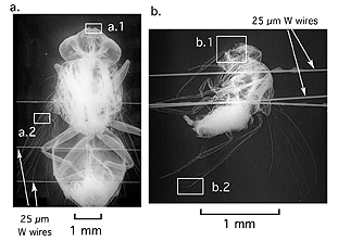
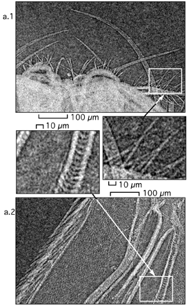
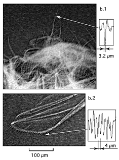
The first images the researchers obtained were fuzzy blobs. But as the technique has been refined, the images have become much sharper. Recently developed radiographs of a dead housefly, collected from the floor of the laboratory, clearly show fine structures, such as hairs, only a few microns across.
As with traditional X-rays, it is also possible to take X-pinch radiographs of living organisms. Hammer and his colleagues have succeeded in making radiographs of a live ant -- however, they had difficulties getting the ant to pose for the picture. "The ant walks around in this little chamber, and so you don't know what angle you're going to X-ray it at. It's much easier to do a dead fly," says Hammer.
The Cornell plasma lab soon will begin collaboration with scientists in the Cornell College of Veterinary Medicine to determine whether this new imaging technology could have important applications for medicine or biology.
This release was prepared by Cornell graduate student Lissa Harris.
Media Contact
Get Cornell news delivered right to your inbox.
Subscribe