The physician as computer scientist: Skorton and medical imaging
By Bill Steele
One part of David Skorton's career echoes a recent major theme in Cornell scholarship: the application of computer technology and computer science ideas in other disciplines.
For many years Cornell has pursued a "strategic initiative" in computing and has created a college-level Faculty of Computing and Information Science (CIS) to recognize that computing is now an important tool in almost every area of research and scholarship. And for many years, Skorton has worked to apply computer processing to medical imaging.
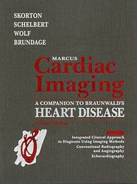
As a cardiologist, he has been particularly interested in imaging of the heart. Quietly buried in his long résumé is his role as editor in chief of and a major contributor to what may be the definitive reference, Marcus Cardiac Imaging (W.D. Saunders, 1996). His work in this field led to his joint appointment to the faculties of medicine and electrical and computer engineering at the University of Iowa. At Cornell he will hold faculty positions in Internal Medicine at Weill Cornell Medical College and in Biomedical Engineering on the Ithaca campus.
Physicians have long depended on X-rays, computerized tomography (CT) scans, magnetic resonance imaging (MRI) and echocardiograms (ultrasound images). Skorton has researched ways to use computers to pull quantitative information out of those images.
Computer "vision" that recognizes the edges of objects or notices differences in texture was first used to analyze photos taken by spy planes, and nowadays helps astronomers scan the sky for fast-spinning neutron stars called pulsars. During his medical residency at the University of California-Los Angeles, Skorton and colleagues applied these techniques to analyze X-rays, echocardiograms and MRI images to determine the dimensions of the heart's chambers, evaluate the condition of tissues, even measure the force with which the left ventricle expels blood into the circulatory system.
By combining a series of "slices" taken by a CT or MRI scan, a computer can generate a three-dimensional image and create a movie simulation of the heart in action. Similar imaging techniques are being applied to view and simulate other organs, to detect tumors and to guide surgeons in their work.
Far more than a theoretical exercise in computer science, these techniques now give physicians an unprecedented amount of information about their patients.
Media Contact
Get Cornell news delivered right to your inbox.
Subscribe