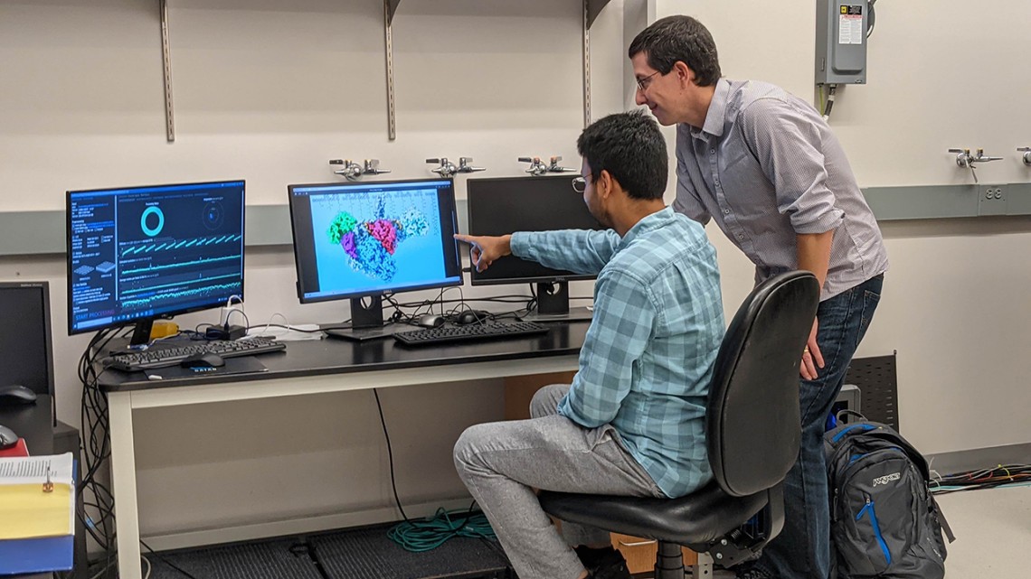
Doctoral student Saket Bagde, left, and Chris Fromme, associate professor of molecular biology and genetics, review the structure of a polyketide synthase enzyme.
Enzyme research unlocks gateway for new medicines
By Krisy Gashler
Cornell scientists have captured a never-before-recorded stage of an antibiotic-producing enzyme’s construction process, opening the gateway for future development of pharmaceuticals, including antibiotics, immunosuppressants and chemotherapeutics.
The study describes the atomic structure of one enzyme – a polyketide synthase – at two different stages of its reaction cycle. This is the first time an entire polyketide synthase has been visualized, said Chris Fromme ’99, one of the study’s authors and associate professor in the Department of Molecular Biology and Genetics. Lead author is Saket Bagde, a doctoral student in Fromme’s lab. The study, “Modular Polyketide Synthase Contains Two Reaction Chambers that Operate Asynchronously,” was published in the Nov. 5 issue of Science.
Many drugs used to treat humans and animals are derived from natural products created by microbes, such as bacteria and fungi. Bagde used the metaphor of an automotive assembly line to describe how enzymes build these natural products. One module would create the car’s frame, then the next would add the doors, and so on. Better understanding this assembly line process can enable scientists to modify those processes to create new drugs.
“If you have antibiotic-resistant bacteria that are able to recognize a drug by its ‘door handle’ and counterattack, you can make another drug that has a different kind of door handle,” Bagde said. “By studying exactly how the reaction is happening, we are paving the way for other scientists to change all of these components. The potential is enormous.”
The most surprising discovery in mapping the synthase was its shape: Two copies of the enzyme associate into a complex with two reaction chambers, so the researchers expected to see the enzyme complex adopt a symmetrical shape. Instead, they discovered that the enzyme complex adopts an asymmetrical shape, which suggests the assembly lines only use one reaction chamber at a time.
“This has far-reaching consequences for drug development, because when you modify the system, if you don’t have in mind how it actually functions, you will not be able to have it function smoothly and you will not get the product that you need,” Bagde said.
That could result in drugs that aren’t as effective as they could be or that cause unnecessary side effects, Fromme said.
The study authors captured two stages of the assembly line of Lasalocid-A, an antibiotic commonly used to treat livestock, using two different techniques:
- X-ray crystallography, a technique scientists have been using for more than 60 years to understand proteins. Researchers coax proteins into crystallizing and then image them with X-rays to learn more about their structure.
- Cryo-electron microscopy, a much-newer method that enables researchers to freeze molecules and then take high-resolution pictures of them using an electron beam.
“Using these two complementary techniques, we were not only able to confirm our findings, but also we were able to get pictures of the enzyme in two different stages,” Bagde said.
Cornell purchased this cryo-electron microscope in 2018, and the investment “is allowing us to be at the forefront of world leaders in structural biology,” Fromme said.
In addition to Bagde and Fromme, co-authors of the study are Chu-Young Kim ’96, professor of chemistry and biochemistry at the University of Texas at El Paso, and Irimpan Mathews, lead scientist at the SLAC National Accelerator Laboratory at Stanford University. Mathews was a postdoctoral researcher at Cornell from 1996 to 2000.
This research was supported by the National Institutes of Health and the Cornell Center for Materials Research.
Krisy Gashler is a writer for the College of Agriculture and Life Sciences.
Media Contact
Get Cornell news delivered right to your inbox.
Subscribe
