Imaging shows how solar-powered microbes turn CO2 into bioplastic
By David Nutt, Cornell Chronicle
When considering ways to sustainably generate environmentally friendly products, bacteria might not immediately spring to mind.
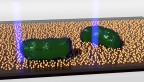
However, in recent years scientists have created microbe-semiconductor biohybrids that merge the biosynthetic power of living systems with the ability of semiconductors to harvest light. These microorganisms use solar energy to convert carbon dioxide into value-added chemical products, such as bioplastics and biofuels. But how that energy transport occurs in such a tiny, complex system, and whether the process can be improved, is still unclear.
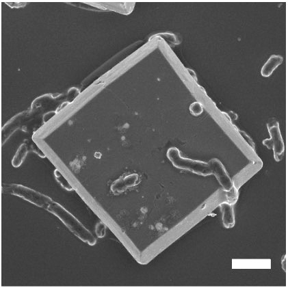
A scanning electron microscopy image shows bacterium Ralstonia eutropha cells on top of a bismuth vanadate semiconductor.
Cornell researchers have developed a multimodal platform to image these biohybrids with single-cell resolution, to better understand how they function and how they can be optimized for more efficient energy conversion.
The team’s paper, “Single-Cell Multimodal Imaging Uncovers Energy Conversion Pathways in Biohybrids,” published July 27 in Nature Chemistry. The co-lead authors are postdoctoral researcher Bing Fu and former postdoctoral researcher Xianwen Mao.
The project was led by Peng Chen, the Peter J.W. Debye Professor of Chemistry in the College of Arts and Sciences. The effort is an offshoot of a larger collaboration – with Tobias Hanrath, professor at the Smith School of Chemical and Biomolecular Engineering in Cornell Engineering, and Buz Barstow, Ph.D. ’09, assistant professor of biological and environmental engineering in the College of Agriculture and Life Sciences – that was funded by the U.S. Department of Energy (DOE) to explore microscopic imaging of microbes as a way to advance bioenergy research.
Biohybrid research has typically been conducted with bacteria in bulk – essentially a large amount of cells in a bucket, Peng said – emphasizing the overall yield of the value-added chemicals and the collective behaviors of the cells, rather than the underlying mechanism that enables the complex chemical transformation.
“Biology is very heterogeneous. The individual cells are very different. Now, in order to interrogate it better, you really need to measure it at a single-cell level,” Chen said. “This is where we come in. We provide quantitative assessments of protein behaviors and also a mechanistic understanding of how the electron transport occurs from the semiconductor to the bacteria cell.”
The new platform combined multi-channel fluorescence imaging with photoelectrochemical current mapping to survey the bacterium Ralstonia eutropha. The platform was able to simultaneously image, track and quantitate multiple proteins in the cell while also measuring the flow of electrons, ultimately correlating the cellular protein properties and electron transport processes.
The researchers successfully differentiated the functional roles of two types of hydrogenases – one bound to the cell’s membrane, and a soluble one in the cytoplasm – that help metabolize hydrogen and drive CO2 fixation. While the soluble hydrogenase is known to be critical for metabolizing hydrogen, the researchers found that the membrane-bound hydrogenase, while less important, actually facilitates the process and makes it more efficient.
In addition, the researchers obtained the first experimental evidence that the bacteria can uptake a large amount of electrons from semiconductor photocatalysts. The team measured the electron current and found it be three orders of magnitude larger than what scientists previously thought, which suggests that future bacteria strains could be engineered to improve the efficiency of energy conversion.
The researchers also discovered that membrane-bound and soluble hydrogenases play an important role in mediating the electron transport from the semiconductor into the cell. Meanwhile, not only can the cell accept electrons; it can also spit them out in the opposite direction, without the assistance of hydrogenases.
The imaging platform is generalizable enough that it can be used to study other biological-inorganic systems, including yeast, and for other processes, such as nitrogen fixation and pollutant removal.
“Our multimodal imaging platform is powerful, but it of course has its own limits,” Chen said. “We can image and study proteins, but our approach does not allow us to analyze small molecule compositions. And so one can think about further integrating our approach with other techniques – for example, nanoscale mass spectrometry – so it would be really powerful. We’re not there yet.”
Co-authors include Hanrath and Barstow; postdoctoral researcher Youngchan Park; doctoral students Zhiheng Zhao, Tianlei Yan, Farshid Salimijazi, and Mokshin Suri; Danielle H. Francis, M.S. ’17; Won Jung, Ph.D. ’18; former postdoctoral researcher Wenjie Li; and laboratory technician Brooke Pian ’13.
The research was supported by the DOE’s Biomolecular Characterization and Imaging Science program.
The researchers made use of the Cornell Center for Materials Research Shared Facilities, which is supported through the National Science Foundation’s MRSEC program.
Media Contact
Get Cornell news delivered right to your inbox.
Subscribe