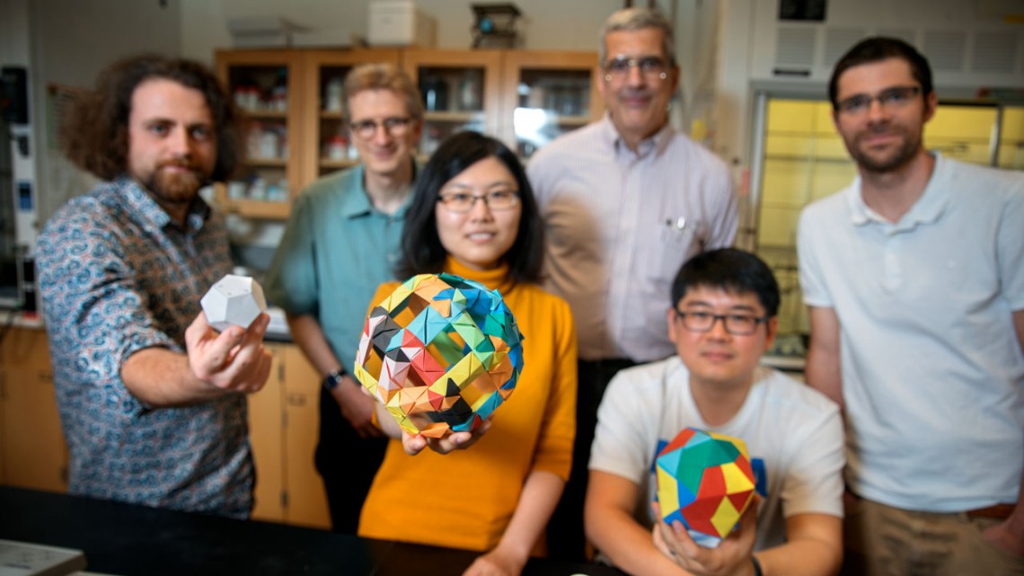
Doctoral student Melik Turker, left, holds a model of a dodecahedron in the lab of Ulrich Wiesner. Also pictured are doctoral student Yunye Gong, center, holding a model of a cage structure, and postdoctoral researcher Kai Ma, holding an icosahedron. The group's paper on their discovery of nanoscale 12-sided silicon cage structures published recently in Nature; in the back row, left-to-right, are Wiesner, engineering professor Peter Doerschuk and postdoc Tangi Aubert.
Collaboration yields discovery of 12-sided silica cages
By Tom Fleischman
What do you call a materials science discovery that was given a major boost by a lecture from a Nobel laureate in chemistry, used cryogenic electron microscopy (cryo-EM), and was pushed further along by a doctoral student’s thesis on machine learning?
Typical Cornell research.
In a paper published in Nature, a team led by Uli Wiesner, the Spencer T. Olin Professor of Engineering in the Department of Materials Science and Engineering, reports discovery of 10-nanometer, individual, self-assembled dodecahedral structures – 12-sided silica cages that could have applications in mesoscale material assembly, as well as medical diagnosis and therapeutics.
The team’s paper, “Surfactant Micelle Self-Assembly Directed Highly Symmetric Ultrasmall Inorganic Cages,” was published June 20.
Other members of the team include postdoctoral researchers Kai Ma and Tangi Aubert from the Wiesner Group; Peter Doerschuk, professor in the Department of Electrical and Computer Engineering and in the Meinig School of Biomedical Engineering; and doctoral student Yunye Gong from the Doerschuk Group.
Techniques from Gong’s doctoral thesis, “Computing and Understanding Statistical Models for Heterogeneous Biological Nano-machines,” were used to determine the 3D shape of the cage structures.
“People had suggested that these very complex nanostructures would be structural units of bulk materials,” Wiesner said, “but nobody had ever identified these cages as isolated building blocks.”
The way to achieve that, Wiesner said: arrest the chemical reaction that produces these shapes early on to see the structure at its inception. “This was indeed part of an ongoing effort of silica chemistry optimization in our group,” Ma said.
To image them, particles in water were rapidly frozen to cryogenic temperatures, so rapidly that instead of ice, water becomes a glassy solid. Within the thin glassy films the cages could be imaged at all different orientations by cryo-EM. Approximately 19,000 single-particle images were collected in a large effort by Ma and other members of the Wiesner Group.
Gong’s machine-learning algorithms, originally developed for the study of virus protein cages, were applied to subsets of these images, sorting them into classes and computing a 3D reconstruction for each class. A calculation based on 2,000 images takes about a day.
Much like a hospital CT scan, the single-particle 3D reconstructions revealed the external shape and the internal structure of the particle.
“We were delighted to have the opportunity to collaborate with the Wiesner Group on this problem,” Gong said, “and demonstrate the breadth of what our algorithms and software can do.”
The group says this might be the first time that single-particle 3D reconstruction of cryo-EM images using artificial intelligence – a rapidly developing technique in structural biology – has been successfully applied to synthetic materials discovery.
“When I first raised the idea of confirming the cage structure by this technique, most people did not believe this was possible because of the material’s complexity,” Ma said.
“That this revealed a beautiful dodecahedron-type cage, the most highly symmetric of the five Platonic solids already studied in antiquity, was immensely rewarding,” Wiesner said.
So where does the Nobel laureate fit in? Several years back, Wiesner sat in on a lecture by Roald Hoffmann, the Frank H.T. Rhodes Professor Emeritus in the Department of Chemistry and Chemical Biology. One topic of the lecture was so-called clathrate cage structures. A clathrate is a compound in which a guest molecule is trapped within the crystal cage of another.
That got Wiesner thinking about the work his lab had been doing on nanoscale structures, which had appeared as just rings in preliminary examination. “After the lecture, I literally ran over to Duffield Hall to tell Kai Ma that I thought the higher-order structures he was sometimes seeing would probably be clathrate-type structures,” Wiesner said. “Kai did more microscopy work and said, ‘Uli, I think you’re right.’”
That serendipitous connection, and the unlikely collaboration involving machine learning with Doerschuk and Gong, is par for the course at Cornell, Wiesner said.
“It’s a typical Cornell story,” he said. “You meet these people from different departments, and everything contributes to a major discovery in science of a beautiful structure that had never been seen before.”
Doerschuk concurred: “I came to Cornell in 2006. One of the main attractions was the claim that barriers between departments are low and that interdisciplinary work is encouraged. Having now worked at Cornell for a decade, I’m delighted to report that the claim is true.”
In the paper, Wiesner contends that “based on recent successes … of ultrasmall fluorescent silica nanoparticles [“Cornell dots”] … a whole range of novel diagnostic and therapeutic probes with drugs hidden inside of the cages can be envisaged.”
Doctoral students Teresa Kao and Melik Turker also contributed to this work. Support came from the National Institutes of Health via a U54 Center for Cancer Nanotechnology Excellence grant to the Wiesner group, the National Science Foundation via an award to the Doerschuk group, and Gong’s Google Ph.D. Fellowship in Machine Learning.
The researchers made use of the Cornell Center for Materials Research Shared Facilities, supported by the NSF, and the Nanobiotechnology Center at Cornell.
Media Contact
Get Cornell news delivered right to your inbox.
Subscribe
