Great white work: Scientists renew the study of shark teeth
By Stacey Shackford


The lasting legacy of the great white shark is sharp, strong and pointy: its teeth.
Not only is it the part of the creature that resonates most strongly with people, it's usually the only part left behind after death, as the rest of its skeleton is cartilage.
A team of Cornell scholars is studying living great whites and other sharks as well as fossilized teeth to gain insight into sharks' ancient ancestors.
The microanatomical study of shark teeth has been dormant for decades, but the scientists are renewing it using the latest imaging technology.
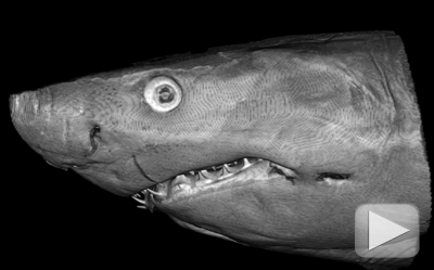
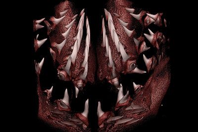
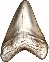
Computed tomography (CT) scans made at the Cornell Micro CT Facility for Imaging and Preclinical Research in Weill Hall enable ecology and evolutionary biology professor Willy Bemis, graduate student Josh Moyer and Micro CT facilities director Mark Riccio to create detailed high-resolution, three-dimensional models to better analyze tooth anatomy, development and evolution.
"The external anatomy of the teeth is all we've had until these CT scans," Bemis said.
Scientists can tell a lot from teeth alone, such as what the animal ate and what its life might have been like. But interpreting fossil teeth can be tricky.
Because of a unique regeneration system in which several rows of teeth are continuously grown and rotated every few weeks in a "conveyer belt" manner, it's sometimes hard to tell the age of a great white shark from a single fossilized tooth. Nobody really knows how great white shark teeth develop, and no one has studied their microstructure for 44 years, since Bernhard Peyer wrote an authoritative work on the topic, "Comparative Odontology" in 1968.
"We are trying to answer some pretty basic questions that we were surprised to discover have not been answered," added Moyer, who works in the graduate field of ecology and evolutionary biology. "It's been really fun to go back and look at these well-regarded hallmark papers and apply them to our research, which is cutting edge, and in so doing, bridge the gap."

The new technology allows the team to examine patterns of tissues and vasculature; they can see the channels left behind by blood vessels and capture images of the developing teeth as they begin to mineralize.
Bemis has amassed hundreds of shark jaws and thousands of teeth in his Corson Hall office and at the Cornell University Museum of Vertebrates, some borrowed from collections around the world, and the team will be examining the internal structure of these teeth to see if they offer any clues into fossil teeth.
Bemis also studies living sharks at the Shoals Marine Laboratory, where he is John M. Kingsbury Director. Since 2007 he has a taught a course on shark biology at the lab, which is located on Appledore Island, six miles off the coast of New Hampshire.
Sharks have been around for about 350 million to 400 million years, and there are currently more than 1,100 living species of chondrichthyes, which also include rays, skates and ratfish.
"Sharks are an extremely successful group with a long history of dramatic evolutionary change, not some ancient relic, and they are highly specialized for specific ways of life," Bemis said.
Understanding why they have been so successful throughout their history and evolution could shed light on the evolution of other species as well, he said.
"Sharks are marvelous model organisms for biomedical research," Moyer said. "There's also an inherent savage romance about them that is appealing. We have a beautiful creature with a fascinating natural history that can be appreciated from biological, emotional and artistic perspectives. What's not to like?"
Stacey Shackford is a staff writer at the College of Agriculture and Life Sciences.
Media Contact
Get Cornell news delivered right to your inbox.
Subscribe